Mouse Secondary Antibodies
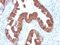
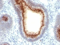
Biotium Cytokeratin 7 (Glandular and Transitional Epithelial Marker) (rOV-TL12/30), CF568 conjugate, 0.1mg/mL
This antibody recognizes an intermediate filament protein (IFP) of 55 kDa, which is identified as cytokeratin 7. This MAb is highly specific to cytokeratin 7 and shows no cross-reaction with other IFPs. Cytokeratin 7 is a basic cytokeratin, which is found in most glandular and transitional epithelia but not in the stratified squamous epithelia. Keratin 7 is expressed in the epithelial cells of ovary, lung, and breast but not of colon, prostate, or gastrointestinal tract. This MAb is highly useful in distinguishing ovarian carcinomas (keratin 7 ) from colon carcinomas (keratin 7-).Primary antibodies are available purified, or with a selection of fluorescent CF Dyes and other labels. CF Dyes offer exceptional brightness and photostability. Note: Conjugates of blue fluorescent dyes like CF405S and CF405M are not recommended for detecting low abundance targets, because blue dyes have lower fluorescence and can give higher non-specific background than other dye colors.


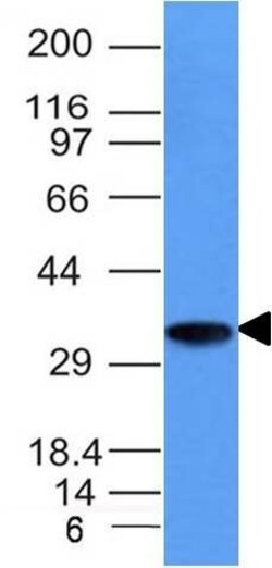
Biotium TDP2 / EAPII (TDP2/1258), CF488A conjugate, 0.1mg/mL
This MAb recognizes a protein of 41 kDa, which is identified as TDP2. It is a member of a superfamily of divalent cation-dependent phosphodiesterases. The encoded protein associates with CD40, tumor necrosis factor (TNF) receptor-75 and TNF receptor associated factors (TRAFs), and inhibits nuclear factor-kappa-B activation. This protein has sequence and structural similarities with APE1 endonuclease, which is involved in both DNA repair and the activation of transcription factors. DNA repair enzyme that can remove a variety of covalent adducts from DNA through hydrolysis of a 5'-phosphodiester bond, giving rise to DNA with a free 5' phosphate. Catalyzes the hydrolysis of dead-end complexes between DNA and the topoisomerase 2 (TOP2) active site tyrosine residue. Hydrolyzes 5'-phosphoglycolates on protruding 5' ends on DNA double-strand breaks (DSBs) due to DNA damage by radiation and free radicals. The 5'-tyrosyl DNA phosphodiesterase activity can enable the repair of TOP2-ind
Biotium Cytokeratin 15 (Esophageal Squamous Cell Carcinoma Marker)(KRT15/2554), 0.2mg/mL
Keratin 15 is a type I keratin which is expressed only in basal keratinocytes in stratified epithelia and does not appear to have a natural type II expression partner. Keratin 15 is down regulated in activated keratinocytes. Cytokeratin 15 is a specific marker of stem cells of the hair-follicle bulge and may be a useful marker for diagnosis between basal cell carcinoma (BCC) and trichoepithelioma. Trichoblastoma are benign neoplasms of follicular differentiation frequently found in nevus sebaceous. Many morphologic features are shared with nodular basal cell carcinoma, sometimes rendering a diagnosis difficult. Trichoblastoma and BCC show variable expression of Cytokeratin 15 and Cytokeratin 19, and absence of hair keratins. Primary antibodies are available purified, or with a selection of fluorescent CF Dyes and other labels. CF Dyes offer exceptional brightness and photostability. Note: Conjugates of blue fluorescent dyes like CF405S and CF405M are not recommended for
Biotium Human IgG Immunoglobulin(B33/20), CF740 conjugate, 0.1mg/mL
Recognizes a protein of 75 kDa, identified as γ heavy chain of human immunoglobulins. Its epitope maps in CH2 domain of Fc region of IgG. It reacts with all sub-classes of γ chain of human immunoglobulins. It does not cross-react with α (IgA), μ (IgM), ε (IgE), or δ (IgD), heavy chains, T-cells, monocytes, granulocytes, or erythrocytes. This MAb is useful in the identification of leukemias, plasmacytomas, and certain non-Hodgkin's lymphomas. The most common feature of these malignancies is the restricted expression of a single heavy chain class. Demonstration of clonality in lymphoid infiltrates indicates that the infiltrate is clonal and therefore malignant.Primary antibodies are available purified, or with a selection of fluorescent CF Dyes and other labels. CF Dyes offer exceptional brightness and photostability. Note: Conjugates of blue fluorescent dyes like CF405S and CF405M are not recommended for detecting low abundance targets, because blue dyes have lo
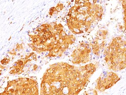
Biotium Arginase 1 (Hepatocellular Carcinoma Marker)(ARG1/1125+ ARG1/1126), 0.2mg/mL
This antibody recognizes a protein of 35-38 kDa, which is identified as Arginase 1 (ARG1). Arginase is a manganese metallo-enzyme that catalyzes the hydrolysis of arginine to generate ornithine and urea. Arginase I and II are isoenzymes which differ in subcellular localization, regulation, and possibly function. Arginase I is a cytosolic enzyme, which is expressed mainly in the liver as part of the urea cycle, whereas arginase II is a mitochondrial protein found in a variety of tissues. Antibodies to Arginase 1 label hepatocytes in normal tissues and granulocytes in peripheral blood. Arginase 1 is a sensitive and specific marker for identification of hepatocellular carcinoma.Primary antibodies are available purified, or with a selection of fluorescent CF Dyes and other labels. CF Dyes offer exceptional brightness and photostability. Note: Conjugates of blue fluorescent dyes like CF405S and CF405M are not recommended for detecting low abundance targets, because blue dyes
Biotium CD74(CLIP/1133), CF740 conjugate, 0.1mg/mL
Recognizes proteins of 33, 35 and 41 kDa, which are identified as various isoforms of CD74. Its epitope is localized in the extracellular domain of CD74. CD74 is a type II transmembrane protein which binds to the peptide binding groove of newly synthesized MHC class II alpha/beta heterodimers and prevents their premature association with endogenous polypeptides. The CD74 molecule plays a critical role in the presentation of peptides, by the MHC class II antigens, to CD4 positive lymphocytes. CD74 is expressed on MHC class II positive cells including B cells, a subset of activated T cells, monocytes, and dendritic cells and by various types of carcinomas. CD74 is expressed primarily by antigen presenting cells, such as B-lymphocytes (from before the pre-B cell stage to before the plasma cell stage), macrophages, and monocytes, and many epithelial cells. Anti-CD74 stains predominantly germinal center lymphocytes and B-cell lymphomas, but rarely T-cell lymphomas. Anti-CD74 has b

Biotium Arginase 1 (Hepatocellular Carcinoma Marker)(ARG1/1125+ ARG1/1126), CF568 conjugate, 0.1mg/mL
This antibody recognizes a protein of 35-38 kDa, which is identified as Arginase 1 (ARG1). Arginase is a manganese metallo-enzyme that catalyzes the hydrolysis of arginine to generate ornithine and urea. Arginase I and II are isoenzymes which differ in subcellular localization, regulation, and possibly function. Arginase I is a cytosolic enzyme, which is expressed mainly in the liver as part of the urea cycle, whereas arginase II is a mitochondrial protein found in a variety of tissues. Antibodies to Arginase 1 label hepatocytes in normal tissues and granulocytes in peripheral blood. Arginase 1 is a sensitive and specific marker for identification of hepatocellular carcinoma.Primary antibodies are available purified, or with a selection of fluorescent CF Dyes and other labels. CF Dyes offer exceptional brightness and photostability. Note: Conjugates of blue fluorescent dyes like CF405S and CF405M are not recommended for detecting low abundance targets, because blue dyes
Biotium Lewis B(2-25LE), CF488A conjugate, 0.1mg/mL
The Lewis histo-blood group system comprises a set of fucosylated glycosphingolipids that are synthesized by exocrine epithelial cells and circulate in body fluids. The glycosphingolipids function in embryogenesis, tissue differentiation, tumor metastasis, inflammation, and bacterial adhesion. They are secondarily absorbed to red blood cells giving rise to their Lewis phenotype. This gene is a member of the fucosyltransferase family, which catalyzes the addition of fucose to precursor polysaccharides in the last step of Lewis antigen biosynthesis. It encodes an enzyme with alpha(1,3)-fucosyltransferase and alpha(1,4)-fucosyltransferase activities. Lewis blood group antigens are carbohydrate moieties structurally integrated in mucous secretions. Lewis antigen system alterations have been described in gastric carcinoma and associated lesions. Anomalous expression of Lewis B antigen has been found in some non-secretory gastric carcinomas and colorectal cancers.Primary antibodies

Biotium Arginase 1 (Hepatocellular Carcinoma Marker)(ARG1/1125+ ARG1/1126), CF740 conjugate, 0.1mg/mL
This antibody recognizes a protein of 35-38 kDa, which is identified as Arginase 1 (ARG1). Arginase is a manganese metallo-enzyme that catalyzes the hydrolysis of arginine to generate ornithine and urea. Arginase I and II are isoenzymes which differ in subcellular localization, regulation, and possibly function. Arginase I is a cytosolic enzyme, which is expressed mainly in the liver as part of the urea cycle, whereas arginase II is a mitochondrial protein found in a variety of tissues. Antibodies to Arginase 1 label hepatocytes in normal tissues and granulocytes in peripheral blood. Arginase 1 is a sensitive and specific marker for identification of hepatocellular carcinoma.Primary antibodies are available purified, or with a selection of fluorescent CF Dyes and other labels. CF Dyes offer exceptional brightness and photostability. Note: Conjugates of blue fluorescent dyes like CF405S and CF405M are not recommended for detecting low abundance targets, because blue dyes
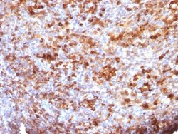
Biotium Neurofilament, phospho (NF-H) (Neuronal Marker)(NE14), Biotin conjugate, 0.1mg/mL
This MAb reacts with a 200 kDa protein, identified as heavy sub-unit of neurofilaments (NF-H). It reacts specifically with the phosphorylated KSP/KEP segment at the C-terminus of the heavy subunit (NF-H) of neurofilaments. After dephosphorylation of neurofilaments with alkaline phosphatase, this Ab no longer binds. Neurofilaments make up the main structural elements of axons and dendrites and are found in neurons, peripheral nerves, and sympathetic ganglion cells. Neurofilaments consist of three major subunits with molecular weights of 68 kDa (NF-L), 160 kDa (NF-M) and 200 kDa (NF-H). Anti-neurofilament stains a number of neural, neuroendocrine, and endocrine tumors. Neuromas, ganglioneuromas, gangliogliomas, ganglioneuroblastomas, and neuroblastomas stain positively for anti-neurofilament. Neurofilaments are also present in paragangliomas as well as adrenal and extra-adrenal pheochromocytomas. Carcinoids, neuroendocrine carcinomas of the skin, and oat cell carcinomas of the
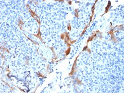
Biotium Napsin A (Lung Adenocarcinoma Marker)(NAPSA/1238 + NAPSA/1239), CF647 conjugate, 0.1mg/mL
Napsin is a pepsin-like aspartic proteinase connected with maturation of surfactant protein B.There are two closely related napsins, napsin A and napsin B. Napsin A is expressed as a single chain protein. Immunohistochemical studies revealed high expression levels of napsin A in human lung and kidney but low expression in spleen. Napsin A is expressed in type II pneumocytes and in adenocarcinomas of lung. The high specificity expression of napsin A in adenocarcinomas of lung is useful to distinguish primary lung adenocarcinomas from adenocarcinomas of other organs.
Biotium CD66 (CEA)(C66/1009), CF568 conjugate, 0.1mg/mL
This antibody recognizes proteins of 80-200 kDa, identified as different members of CEA family. CEA is synthesized during development in the fetal gut and is re-expressed in increased amounts in intestinal carcinomas and several other tumors. This MAb does not react with nonspecific cross-reacting antigen (NCA) and with human polymorphonuclear leucocytes. It shows no reaction with a variety of normal tissues and is suitable for staining of formalin/paraffin tissues. CEA is not found in benign glands, stroma, or malignant prostatic cells. Antibody to CEA is useful in detecting early foci of gastric carcinoma and in distinguishing pulmonary adenocarcinomas (60-70% are CEA positive) from pleural mesotheliomas (rarely or weakly CEA positive). Anti-CEA positivity is seen in adenocarcinomas from the lung, colon, stomach, esophagus, pancreas, gallbadder, urachus, salivary gland, ovary, and endocervix.Primary antibodies are available purified, or with a selection of fluorescent CF
Biotium Cytokeratin 16 (KRT16) (Suprabasal Keratinocyte Marker)(KRT16/1714), CF740 conjugate, 0.1mg/mL
Cytokeratins are a family of intermediate filament proteins that assemble into filaments through forming heterodimers of one type I Cytokeratins (Cytokeratins 9 to 23) and one type II Cytokeratins (keratins 1 to 8). The cytokeratin proteins play a critical role in differentiation, as well as tissue specialization and function, to maintain the overall structural integrity of epithelial cells. Cytokeratins are also useful markers in identifying the origin of metastatic tumors. Cytokeratin 16 is expressed in benign stratified squamous epithelium and squamous cell carcinoma of the head and neck, as well as luminal cells of mammary gland and sweat ducts. It is absent in non-invasive breast carcinomas and normal breast tissue. Primary antibodies are available purified, or with a selection of fluorescent CF Dyes and other labels. CF Dyes offer exceptional brightness and photostability. Note: Conjugates of blue fluorescent dyes like CF405S and CF405M are not recommended for dete
Biotium Cytokeratin 20 (KRT20) (Colorectal Epithelial Marker)(KRT20/1993), CF488A conjugate, 0.1mg/mL
This MAb recognizes an intermediate filament protein of 46 kDa, identified as cytokeratin 20 (KRT20). KRT is abundantly expressed in goblet cells and enterocytes of the gastrointestinal tract. It is a useful marker of pancreatic and colorectal cancer. KRT20 is expressed under normal, hyperplastic and neoplastic conditions. It has been detected in adenocarcinomas of the colon, stomachand biliary tract. Breast carcinomas are generally non-reactive. Primary antibodies are available purified, or with a selection of fluorescent CF Dyes and other labels. CF Dyes offer exceptional brightness and photostability. Note: Conjugates of blue fluorescent dyes like CF405S and CF405M are not recommended for detecting low abundance targets, because blue dyes have lower fluorescence and can give higher non-specific background than other dye colors.